Call 919-684-7777 or fax 919-668-1221.
A 63-year-old male patient with a history of multiple sclerosis presented to the Duke Health ER with unbearable bilateral hip/sacral pain. The pain persisted for a few months, but had progressed significantly over the last two weeks. As a cattle farmer in rural North Carolina, the pain was preventing his ability to work and function, leaving him barely able to walk. “There was a closer hospital, but I chose to drive the 100 miles to Duke,” says the patient.
After arriving at the ER, a CAT scan revealed a very large calcified synovial cyst in the epidural space, occluding his spinal canal and causing nerve compression. Oren Gottfried, MD, spine surgeon, reviewed his imaging and determined surgery was required to remove the cyst.
“This was the second largest synovial cyst I have ever seen,” said Gottfried. “Due to its calcification and size, taking out the cyst could affect the nerves and pose catastrophic risks of paralysis to the patient.”
The patient was discharged, and surgical planning proceeded. An MRI was examined for nerve anatomy and compression. “The cyst was so large and calcified that it occluded the nerves on the imaging—you could only see the nerves above and below,” explains Gottfried.
Within a couple of weeks, surgery was scheduled. “Dr. Gottfried stood out,” says the patient. “It was pretty grim; I was told they were going to try to make sure I could control my bladder and bowels after surgery, and paralysis was possible if we didn’t act soon, but I told Dr. Gottfried I knew he could do better than that.”
How did Gottfried approach the high-risk surgery to resolve pain and restore the patient’s function?
For the procedure, Gottfried did a L4-5 laminectomy, drilling the normal bone away to access the cyst. He found a plane above and below the cyst, which was very hard and calcified. “I was able to expose the cyst, but it was very stuck to the nerves. I slowly started resecting small parts of it, millimeter-by-millimeter to clean it out and finally remove the wall of the cyst that was stuck to the nerves,” he adds.
Gottfried explains that for many patients, large cysts or the surgery to treat them can cause spinal instability. “The degeneration of the spine causing these cysts is so disruptive that, sometimes, it can leave the spine unstable. We do our best to avoid fusion, though, and I was very happy that we were able to maintain his spine stability and avoid fusion in this case.”
Gottfried recounts that he could see the nerve compression releasing immediately after removing the cyst: “When I took out the lesion completely, I saw the nerve sack expand, and the nerves responded in real time. I knew we did the job.”
The patient recalls waking up from anesthesia after surgery and knowing right away he was healed. “I had pain from the surgery, but the original pain I felt from the cyst was gone,” says the patient.
After a few weeks of healing, the patient returned to the farm to continue the cattle work his family has done for two generations. “I couldn’t have asked for a better outcome,” he says.
Gottfried performs a high volume of spinal cyst consultations and surgeries. “We personalize each patient’s plan; not all patients need surgery,” he says. “Some spinal cysts can be managed without surgery if they aren’t compressing nerves and causing functional deficits.”
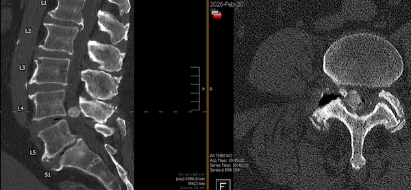
Imaging showing large spinal cyst pre-surgery
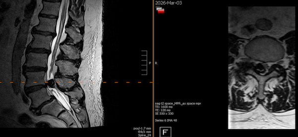
Imaging showing complete cyst removal post-surgery