T-cell lymphopenia is a prominent characteristic of glioblastoma (GBM). However, the cause is not well understood. Now, Duke researchers have found the missing T-cells. In patients with GBM and other intracranial tumors, naïve T-cells are sequestered in large numbers in the bone marrow, trapped there because of tumor-imposed loss of a surface receptor required for egress.
Published in August 2018 in Nature Medicine, the findings suggest that a new approach to immunotherapy, one that restores T-cell egress from the bone marrow, may be a way to grant efficacy to T-cell–based cancer immunotherapies that are currently ineffective.
“Part of the problem with all these immunotherapies—particularly for glioblastoma and other tumors that have spread to the brain—is that the immune system is shot,” says lead author, Peter Fecci, MD, PhD, director of the Brain Tumor Immunotherapy Program in Duke Neurosurgery. “If the goal is to activate the T-cells and the T-cells aren’t there, you’re simply delivering therapy into a black hole.”
Previously, the lymphopenia observed in patients with GBM was often attributed to treatment. But when the researchers reviewed patient records, they found that the dramatic T-cell depletion, comparable to people with AIDS (CD4 T-cell counts < 200 l-1), was also present in treatment-naïve patients. They began to search for the “missing” T-cells.
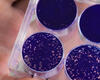
Given that splenomegaly and splenic T-cell sequestration is common in a number of disease states, the researchers first examined the spleen. But the spleens of patients with GBM were abnormally small, as were the thymus and cervical lymph nodes. The number of naïve T-cells in particular was reduced, suggesting that T-cell production in the bone marrow was somehow impaired in these patients. Instead, when they turned to the bone marrow, they found that T-cell numbers were significantly increased.
“It’s totally bizarre—this is not seen in any disease state,” Fecci says. “This appears to be a mechanism that the brain possesses for keeping T cells out, but it’s being usurped by tumors to limit the immune system’s ability to attack them.”
In searching for a mechanism and potential therapeutic targets, Fecci and colleagues found that the sequestered T cells lacked surface expression of sphinogsine-1-phosphate receptor 1 (S1P1), a G-coupled protein receptor (GPCR) that plays an important role in lymphocyte trafficking.
Furthermore, loss of S1P1 surface expression was sufficient to cause T-cell sequestration in the bone marrow. On the flip side, stabilization of S1P1 surface expression enabled T cells to resist sequestration and travel to the tumor, indicating that the process is reversible, Fecci says.
Stabilization of S1P1 T cell surface expression alone was not sufficient to improve survival in a mouse model. However, when coupled with a T-cell activating therapy, median survival was prolonged, suggesting that the new approach may hold promise as a therapeutic adjunct.
At present, no therapies that stabilize S1P1 expression exist. Fecci and colleagues are collaborating with Duke scientist Robert Lefkowitz, MD, whose 2012 Nobel Prize in Chemistry honored his work with GPCRs, to develop drugs that can restore surface expression.
“We are hopeful that this finding provides a missing element that would enable more immunotherapies to be effective for more people,” Fecci says. He said the finding could also work in reverse, offering a new approach to quell autoimmune disorders by activating T-cell sequestration.