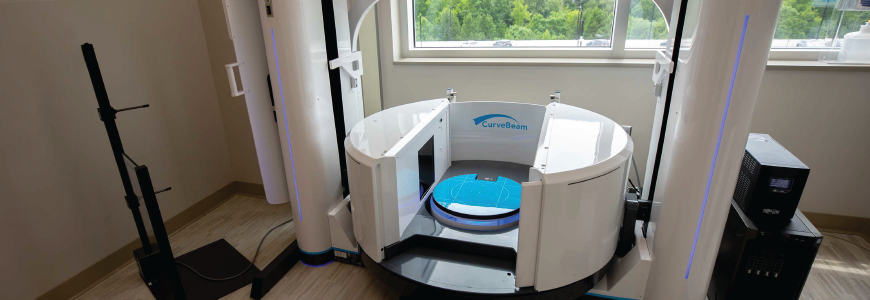
New scanning technology that allows three-dimensional imaging of the foot and ankle under weight-bearing conditions has transformed the care of several conditions, according to Cesar de Cesar Netto, MD, PhD, an orthopaedic surgeon who joined Duke’s Division of Foot and Ankle in May.
Duke is one of a few centers in North Carolina to offer these cone-beam CT scanners, which create 3D images while exposing the patient to one-sixth to one-tenth of the radiation of the traditional CT scans taken while a patient is lying down. “A cone of X-ray rotates around the body once in a single pass for bilateral foot and ankle imaging that takes only about 25 seconds,” de Cesar Netto says.
Although the technology is relatively new, de Cesar Netto says that it should be considered the standard of care for many conditions. “Flatfoot, syndesmotic ankle injuries or high ankle sprains, and hallux valgus or bunions are the three treatments that weight-bearing CT scanning has changed the most,” he says.
“For lower extremity orthopaedic pathologies, the standing body weight load is paramount in the assessment and is when the pathologies show up,” de Cesar Netto says. “For example, a foot without weight on it may have a normal appearance, but when you put body weight load on it, the foot collapses.”
“Similarly, when you put stress on the syndesmotic joint, you can see the instability that you were not able to see before,” de Cesar Netto says. “Under load is how our lower extremity joints work, so judging alignment or getting measurements out of non-weight-bearing imaging doesn’t make any sense.”
Radiographs can be taken under weight-bearing conditions, but do not provide a complete picture. “Foot and ankle problems are complex and three-dimensional, but with conventional X-rays we were seeing them in only two dimensions,” de Cesar Netto says.
“With the data that has come out of three-dimensional, weight-bearing CT imaging, our understanding of flatfoot issues was re-invented, and we are changing treatment based on this new understanding. Currently, it would not be too bold to say that treating a collapsed flatfoot without weight-bearing imaging is doing a disservice to the patient,” he says.
Two new scanners are available at Duke Orthopaedics Arringdon, one for imaging the entire lower extremity from the hips down and one for foot and ankle only.