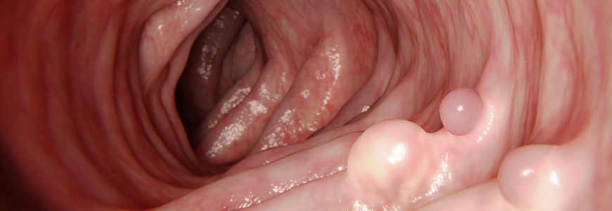
Complete removal of large gastrointestinal (GI) tract polyps is often challenging but essential in reducing the risks for colorectal cancer, particularly in light of evidence that up to 30% of patients who have had endoscopic mucosal resection (EMR) of colon polyps have recurrence of adenomatous tissue at the removal site.
Duke gastroenterologists, including Joshua P. Spaete, MD, are using novel tools and advanced endoscopic techniques that offer an array of solutions for approaching polyp removal, addressing incomplete resection, and sparing patients from unnecessary surgery. The novel techniques include:
Endoscopic Submucosal Dissection
Duke is one of only a few centers in the Southeast offering endoscopic submucosal dissection (ESD), an advanced and technically demanding procedure that allows complete removal of a polyp in one piece. Fluid is injected into the submucosa of the gastrointestinal (GI) tract, creating a cushion of fluid that separates the inner and outer layers of the organ wall. An electrosurgical knife is used to cut a circle around the lesion, and the gastroenterologist slowly works the knife underneath the lesion to remove it in one piece. Spaete and colleagues Darin L. Dufault, MD, and M. Stanley Branch, MD, are among a small number of specialists in the U.S. with significant experience performing ESD. This technique can be used for previously treated polyps but can also be used to remove large polyps at the time of initial evaluation. Resection of a polyp in one piece significantly decreases the likelihood of recurrent polyp formation that may need to be treated in the future.
Snare Tip Soft Coagulation
While complete removal of all polyp tissue at the time of initial EMR is the goal, recurrent polyp can form in up to one-third of all large EMR cases. One technique to address this is treatment of the resection margin with soft coagulation at the time of EMR of large polyps. “Treating the post-EMR margin with snare tip soft coagulation can reduce recurrence of polyps from up to 30% down to 5%,” says Spaete. The procedure uses a low-voltage limited coagulation; traditional cautery uses higher voltages which may unnecessarily damage tissue.
Double-balloon Enteroscopy
Duke is one of the few centers in the Southeast that offer this minimally invasive procedure, which allows gastroenterologists to reach and treat colon polyps or areas of bleeding in the GI tract that are difficult to access. The system has an endoscope and an overtube with two attached balloons, which inflate and deflate to grip and compress the walls of the small intestine and allow the scope to advance deep into the small bowel. This allows access to areas of the small bowel that could not otherwise be reached endoscopically. Interventions for bleeding lesions and the removal of polyps deep within the small bowel, which previously would require surgical exploration and intervention, may now be performed endoscopically.
Full-thickness Resection Device
One novel tool that Duke utilizes is the FTRD System (Ovesco, Tuebingen, Germany), a full-thickness resection device which has an applicator cap and deployable clip mounted on the endoscope with a snare that’s used to cut tissue. The clip is deployed at the targeted site and all tissue between the clip and the endoscope can be removed with the snare. The clip locks to itself to prevent a perforation within the bowel wall at the removal site. This can be used for any lesion within the stomach, small bowel or colon that can be accessed with the device and is particularly useful for polyps within the colon that have previously been treated and polyp tissue remains. “The device allows us to go all the way through the deepest muscle layer, remove all the scar tissue, and make sure the deeper section of the polyp area is completely clear,” Spaete explains.
EndoRotor Device
Another novel tool is the EndoRotor device (Interscope, Whitinsville, MA), which can be used to treat refractory polyps. It uses a rotating cutting blade that shaves off all the layers of polyp tissue above the muscle layer. The endoscope is advanced to the area of the polyp, epinephrine is injected, and the device is passed through the working channel of the scope to the area of concern. Several passes are made with the device until removal of the polyp tissue is achieved. This has been effective in treating polyps that may have significant scarring from prior endoscopic therapy.
Hot Biopsy Forceps
Although it’s not a new device, and its use had previously fallen out of favor for the treatment of small polyps, Spaete says Duke gastroenterologists are using hot biopsy forceps to treat small areas of recurrent or residual polyp tissue that are significantly fibrosed and cannot be adequately lifted. Using the avulsion technique, the area of concern is grasped and moved away from the underlying layers, and electricity is applied to the forceps to simultaneously remove and electrocoagulate the tissue. “This technique is effective when there’s a small amount of scarred polyp tissue remaining after a polyp has been removed in pieces,” he explains.
Tips for Treating Polyps
Spaete encourages gastroenterologists to keep these tips in mind when treating patients with polyps:
- For large polyps that cannot easily be removed and may be difficult for another specialist to locate on follow-up, tattoos can be used but it’s best to place them in a site that is not underneath the polyp. Tattoos placed beneath a polyp can lead to fibrosis and limit the ability to fully remove it in the future. One tattoo, approximately 3 cm downstream on the opposite wall, should allow for accurate identification on follow-up and limit scarring.
- For polyps that are of questionable concern, biopsies should be taken but the number of biopsies should be limited to prevent scarring, which hinders the ability to remove polyps later via endoscopic resection.
- For refractory polyp tissue found after previous piecemeal removal, patients should be referred to Duke Gastroenterology as soon as possible for the best chance at removing it permanently and preventing the patient from having numerous repeat colonoscopies and procedures.
