For patients diagnosed with a rare but aggressive form of bone cancer, surgical resection and reconstruction of the affected bone or soft tissue can lead to a loss of joint stability with a prosthesis or a limb amputation. However, a 3D-printing technology featuring a Duke Engineering innovation helps Duke sarcoma specialists to create a more integrated implant solution that helps patients better preserve long-term function and improve chances for a positive outcome.
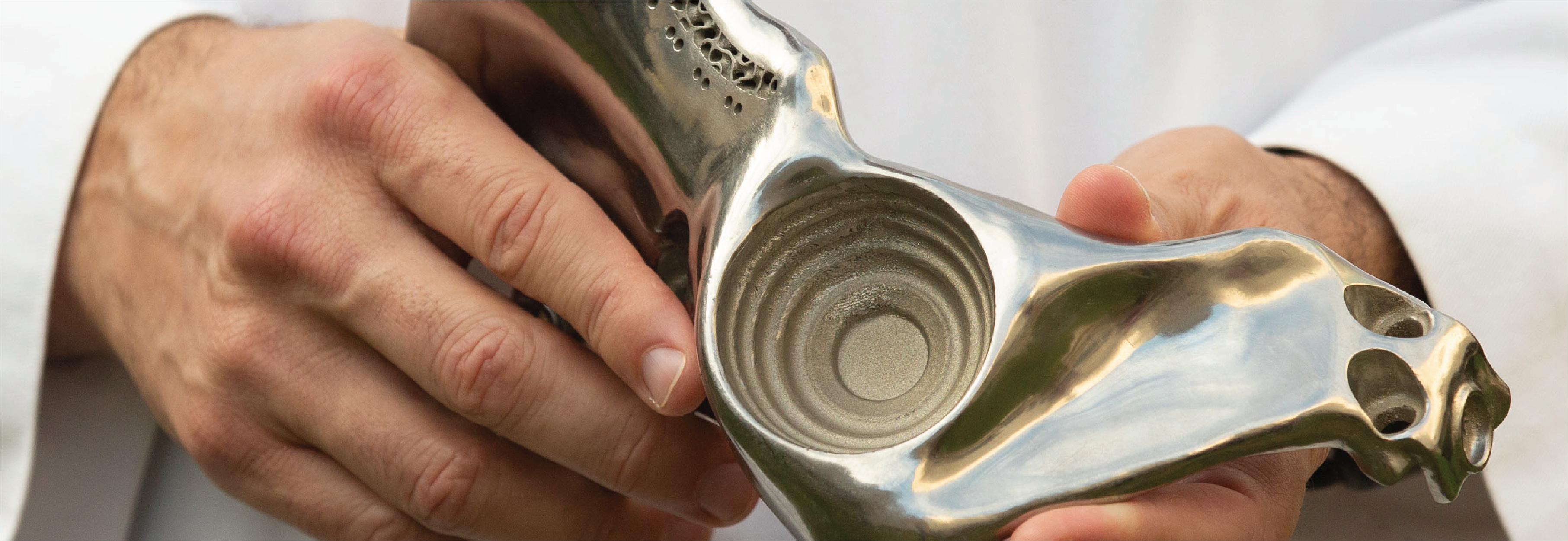
Custom 3D printing is standard practice at Duke, but the implants designed and printed by Duke’s Division of Orthopaedic Oncology are unique in their inclusion of a gyroid—a mathematical shape that allows for the maximum amount of surface area in a unit of volume. (See the small grooves along the edge of the implant in Figure 1.)

This detail is significant because it allows the most bone possible to grow into the structure, helping the remaining bone or cartilage to grow together with the implant. (The implant design innovation originated with Ken Gall, MD, PhD, a Duke Engineering professor who co-founded Restor3d (Durham, NC), which prints the materials).
Using CT scans and precision software, Duke sarcoma specialists can 3D print the piece of bone needed for the implant as well as the cutting guides used during the reconstruction.
Duke’s three orthopaedic surgical oncologists Brian Brigman, MD, PhD, Will Eward, MD, DVM, and Julia Visgauss, MD have deep expertise in treating these lethal sarcomas, which require extensive case review, multidisciplinary input, and a tailored approach for each patient. Additionally, the Duke Sarcoma Center was recently established as a center of excellence for sarcoma treatment and research, and the disease group at Duke has been recognized for its innovative personalized treatment grounded in state-of-the-art clinical and basic-science research.
“In addition to surgical margin, one of the biggest factors that makes an impact on surgical outcomes is being treated at a highly specialized institution with a high volume of these patients, and Duke has one of the highest volumes of sarcoma cases anywhere,” Eward says.

Bone tumors are uncommon, with 5,000 to 6,000 cases diagnosed each year in the United States. Although osteosarcoma presents in patients of all ages, it is primarily a disease of children, teenagers, and young adults, while chondrosarcoma occurs most often in people over age 40.
This custom 3D implant solution is applicable to many different parts of the body affected by osteosarcoma or chondrosarcoma—including arms, legs, and chest—but Eward says the pelvis a good example of the full breadth of what this technology enables Duke’s sarcoma specialists to do for patients. (See Figure 2.)
“For long bones like the femur, we can use a standard megaprosthesis to replace the affected piece of the bone or cartilage, but the pelvis is much more difficult to reconstruct because when you resect the bone, there is no longer a socket for the ball of the hip joint,” he explains.

As a result of careful planning, Duke sarcoma specialists 3D print the titanium implant, a model of the residual bone, and cutting guides that are sterilized and used during the up to 10-hour reconstructive surgery. Eward notes that after dissecting essential arteries, veins, and nerves away from the tumor, they would then temporarily affix the cutting guide to the bone, resecting the cancerous area of bone. Finally, surgeons implant a custom prosthesis into the pelvis and secure the socket portion of the hip joint, followed by a hip replacement procedure to complete the pelvic reconstruction. (See Figure 3.)
After surgery, the importance of the gyroid is most evident, Eward says: “When the patient recovers and starts walking around, at some point those screws in the pelvis are going to break, so the gyroid design helps the bone and implant to eventually grow together so that the patient doesn’t rely on those screws for mobility.”
Eward describes Duke’s ability to anatomically reconstruct a patient’s bone or cartilage with a custom 3D-printed implant as “life-changing—it’s literally the difference between having a leg and not having a leg in some cases,” he adds.