Better imaging before hip surgery leads to better outcomes, and Duke is one of the few orthopaedic centers in the country that offers 3-Tesla MRI—a powerful diagnostic tool that avoids painful injections of contrast media in the hip joint and the radiation associated with CT scans—combined with a 3-D modeling program, developed by Duke.
In this system, a single MRI scan provides a wealth of information, giving surgeons a “road map to address the problem” in complex hip surgery, according to Brian Lewis, MD, head of Duke’s Hip Preservation Section.
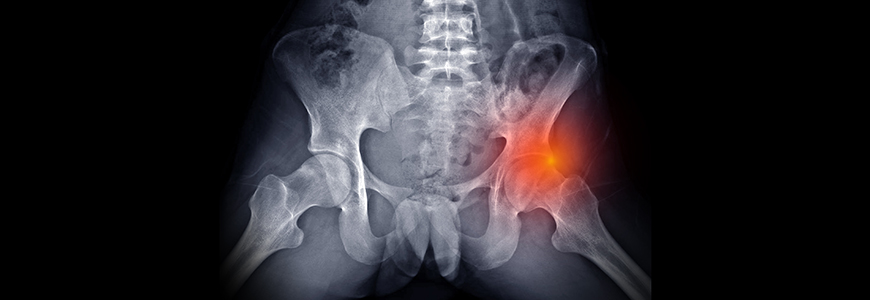
As part of his specialty in hip preservation surgery, Lewis has expertise in addressing hip impingement, a condition in which the femoral head grows into a shape that interferes with smooth movement. A bump in the femoral head can also often damage the labrum.
Hip impingement is a relatively new condition of modern life. Although it is currently present in 30% of the population, the condition is not seen in skeleton remains from hundreds of years ago. It does not have a clear origin. “We think it may be associated with the growth plate in the ball seeing abnormal stress,” Lewis says. The condition seems to develop more commonly from activities like playing goalie in ice hockey.
To refer a patient, call Duke's Consultation and Referral Center at 800-633-3853 or log into Duke MedLink.
Although most cases are asymptomatic, the key diagnostic factor indicating a need for treatment is hip discomfort, such as pain during or after activity, or even pain from long periods of sitting. “If you’re a young person and your hip hurts, there is likely something wrong with the shape of your hip,” Lewis says. “Whether it is dysplasia or impingement, there is something wrong.”
Hip preservation surgery is also relatively new, first offered by Duke in 2012. Typically used in people ages 15 to 50, it relieves symptoms, improves quality of life, and reduces the future need for hip replacement.
The advantage of Duke’s system is that it provides greater resolution in a single scan, with no need for multiple studies. “It allows us to look at all the things that could be wrong with the shape of the hip because we can make a 3-D model. Once we understand what’s wrong with the shape of a hip, we have the best chance of treating it,” Lewis says.
“We use 3-D images prepared ahead of surgery and then use intraoperative x-rays and other advanced tools that help tell us when we have corrected the femur to a more normal shape using a device to reshape the bone,” Lewis says.
In addition to its benefits in hip impingement, Duke’s imaging expertise offers similar treatment advantages for other conditions, such as hip dysplasia. Hip dysplasia often begins in childhood, but pain associated with the condition may not be noticeable until young adulthood. It is the most common cause of hip arthritis in women younger than 50 and becomes harder to treat the longer a patient has had dysplasia.
With hip dysplasia being more common in women than men, patients may appreciate the perspective offered by two new hip dysplasia specialists, Amy Behman, MBChB, PhD, FRCSC, and Elizabeth J. Scott, MD, who recently joined the Duke hip preservation team.