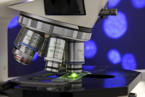
Cancerous Tissue Illuminated During Surgery

According to findings published January 6, 2016, in Science Translational Medicine, a phase 1 trial at Duke Health in 15 study patients undergoing surgery for soft-tissue sarcoma or breast cancer found that cathepsin-activatable Cy5 fluorescent imaging probe LUM015 (Lumicell, Wellesley, MA) identified cancerous tissue and was well tolerated. The imaging technology was developed through collaboration between scientists at Duke, Massachusetts Institute of Technology, and Lumicell.
Reliance on magnetic resonance imaging and computed tomography to guide surgical oncologists in the removal of tumors and tumor margins can sometimes mean that cancerous tissue may go undetected, possibly requiring a second surgery and use of radiotherapy.
“At the time of surgery, a pathologist can examine the tissue for cancer cells at the edge of the tumor using a microscope,” says David Kirsch, MD, PhD, a professor of radiation oncology and pharmacology and cancer biology at Duke and co-senior author of the study. “But, because of the size of the tumor, it’s impossible to review the entire surface during surgery.”
“The goal is to give surgeons a practical and quick technology that allows them to scan the tumor bed during surgery to look for any residual fluorescence,” he explains.
Worldwide, researchers are pursuing techniques to help surgeons better visualize cancerous tissue. The trial results described in the journal suggest that LUM015, the first protease-activated imaging agent for cancer, could prove useful in humans.
The study authors examined both mice and human models, and they discovered that LUM015 accumulated in tumors where it creates fluorescence in cancerous tissue that is, on average, 5 times brighter than that seen in regular muscle. The resulting signals must be detected by a handheld imaging device with a sensitive camera, which Kirsch says Lumicell is also developing.
Brian Brigman, MD, PhD, chief of orthopaedic oncology at Duke and co-senior author on the study, explains that the goal of surgery is to remove 100% of the tumor and resect all of its margins.
“If we can increase the cases where 100% of the tumor is removed, we could prevent subsequent operations and potentially cancer recurrence,” says Brigman. “Knowing where there is residual disease can also guide radiotherapy or reduce how much radiation a patient will receive.”
Researchers at Massachusetts General Hospital are also evaluating the safety and efficacy of LUM015 and the Lumicell imaging device in a prospective study of 50 women with breast cancer. Afterward, Kirsch says other institutions are likely to evaluate whether this technology can be used to decrease the number of patients with breast cancer who need subsequent surgeries.