Blurry Vision in a Young Patient

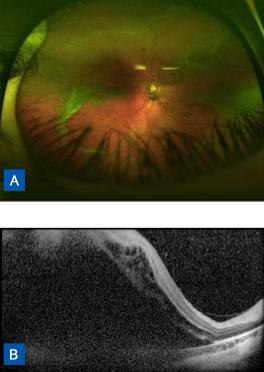
FIGURE 1. (A) Fundus photography of right eye indicating prominent vitreous veils and retinal detachment. (B) Optical coherence tomography shows subretinal fluid extending into macula.
After presenting with blurry vision and reporting the presence of dark spots in her vision, an 8-year-old girl was diagnosed with bilateral retinal detachment and referred to the Duke Pediatric Retina and Optic Nerve Center at the Duke Eye Center (Figure 1). She had a family history of retinal detachment, and her father had type 1 Stickler syndrome.
Question: What steps needed to be taken for this patient in addition to repairing her retinal detachments?

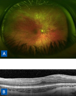
FIGURE 2. (A) Fundus photography shows retina attached in right eye following peripheral laser and scleral buckle. (B) Optical coherence tomography demonstrates recovery of photoreceptors and fair foveal contour with mild epiretinal membranes.
Answer: Because Stickler syndrome is a hereditary condition with sequellae such as retinal detachment for which effective prophylactic interventions are available, the patient and her older brother underwent examination and genetic testing, and, upon confirmation that both children had the syndrome, genetic counseling was recommended.
Stickler syndrome is significantly underdiagnosed, which prevents patients from receiving early interventions that can preserve vision, says Lejla Vajzovic, MD, the Duke pediatric retina specialist who treated the patient. “We need to be thinking about this condition and considering genetic testing any time we see a child with retinal detachment—particularly for a patient like this who is known to have a family member with Stickler syndrome,” she advises.
After performing retinal detachment repair (Figure 2) and collecting blood from the patient, Vajzovic submitted the blood sample for genetic screening. When the test results came back positive for Stickler syndrome, she recommended the patient’s brother be genetically screened and potentially prophylactically treated.
His test results revealed that he also had the disorder, and, in the meantime, subsequent comprehensive eye examination indicated he had developed retinal detachment in 1 eye. Vajzovic repaired the retinal detachment and performed prophylactic cryotherapy in the other eye, a preventive measure that decreases the likelihood of retinal detachment by 7.4-fold, as reported in the Cambridge prophylactic cryotherapy protocol.
Although it was too late to prevent retinal detachment in the original patient, Vajzovic continues to follow her and monitor for retinal tears, which can progress to retinal detachment if left untreated. Now, 2 years later, neither patient has required additional intervention, and both maintain good visual acuity.
“It was really important that we didn’t just repair the patient’s retinal detachment but that we also provided genetic screening and counseling,” Vajzovic remarks. “That allowed us to identify the condition earlier in her brother so we could take some preventive measures. It also enabled us to discuss the probability that the patients’ kids will have the disorder and recommend that they have their kids tested as soon as they are of examinable age.”