A 63-Year-Old Man With Recent-Onset, Debilitating Back Pain
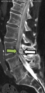
Figure 1. CT Myelogram Sagittal Image. Large calcified mass at L3-L4 (white arrow) with critical spinal stenosis and myelographic block (green arrow); evidence of prior fusion L4-S1
In April 2014, a 63-year-old man presented to the Duke neurosurgery clinic with lower back pain that had begun about 6 weeks earlier. The symptoms had progressed to the point that he was unable to walk more than a few steps without severe pain and weakness. The patient had a remote history of multiple lumbar surgeries: a spinal fusion at L4-S1 in 1993 and several surgeries in 2003, with complications that included infection. Before being referred to Duke, the patient had been extensively evaluated by two other spine surgeons. A CT myelogram performed by one of those surgeons revealed a complete blockage of myelographic dye at L3-L4 by a large mass (Figure 1); however, an earlier MRI from 2005 showed no evidence of this mass.
Duke neurosurgeon Oren Gottfried, MD, examined the patient, who was 6’ 2” and 270 lbs (body mass index of approximately 35 kg/m2), and found that he was unable to stand for more than a few seconds. Lower-extremity strength testing revealed substantial motor weakness in the tibialis anterior and extensor hallucis longus muscles on the left, but no severe strength deficits were present on the right. Patella and Achilles reflexes were absent bilaterally; sensation of the lower extremities appeared to be intact. Surprisingly, the patient reported no bowel or bladder symptoms.
What type of mass do you think was causing this patient’s symptoms?
Answer: A very large ossified synovial cyst.
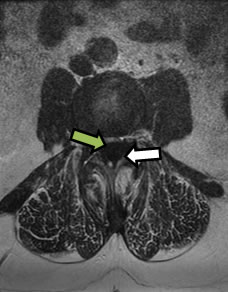
Figure 2. MRI T2-Weighted Axial Image. Large juxtafacet mass at L3-4 from left (white arrow) resulting in severe spinal stenosis on thecal nerve sac (green arrow)
Concerned about the severe stenosis present at L3-L4, Gottfried ordered an MRI to be performed that night (Figure 2), knowing that it would also aid in treatment planning. “The MRI did show what I considered to be the largest cyst I’ve ever seen. His thecal sac was completely obliterated by this cyst,” Gottfried said. “The other interesting thing about this mass was that it was totally calcified—most likely a bony cyst—which was unusual.”
Gottfried suspected that the mass was an ossified synovial cyst, arising from the synovial membrane of the facet joints. Such cysts are usually caused by irritation from arthritis, joint stress from facet arthropathy, and instability. Gottfried explained that synovial cysts are very common as adjacent-level disease in patients who have undergone prior spinal-fusion surgeries.
“The referring neurosurgeon and orthopaedic surgeon thought the mass was an intradural cyst—most likely an arachnoid cyst,” Gottfried said. “But this didn’t really fit with why this patient would have developed it in the first place.” Removal of intradural arachnoid cysts requires very different surgery and management than removal of extradural cysts, Gottfried noted.
Although many spinal cysts can be managed conservatively, the size of the cyst, coupled with the patient’s progressive symptoms, warranted surgical intervention. Two days later, the patient underwent spinal surgery to have the cyst removed. It took Gottfried approximately 1 hour to tease out the adhesions of the ossified cyst using microdissection techniques. “With normal, noncalcified synovial cysts, the incidence of having a dural tear or complication is upwards of 10%,” Gottfried said, “but we were able to remove it without any tearing of the thecal sac and without any injury to the nerves.” To increase stability at the L3-L4 level, Gottfried also performed a spinal fusion. Pathology results showed that the mass consisted of bone only.
The patient woke from surgery with immediate improvement in his pain. “He actually walked postoperatively on day one,” Gottfried said, which was surprising given his preoperative symptoms and the size of the cyst. At 3-month follow-up, the patient was walking and was continuing to regain his endurance. “Considering how much pressure was on the nerves, it will probably take a good 6 months for the patient to get his endurance back,” Gottfried said at that time, adding that he expected the patient to make a full recovery.